
If you have a brain or spinal cord tumour, your healthcare team will create a treatment plan just for you. It will be based on your health and specific information about the cancer. When deciding which treatments to offer for a brain or spinal cord tumour, your healthcare team will consider:
• the type of tumour • the grade of the tumour • the location of the tumour • the size of the tumour • your neurological (nervous system) function • your age and overall health • previous treatmentSurgery is usually used to treat brain and spinal cord tumours. The type of surgery you have depends mainly on the location and size of the tumour. When planning surgery, your healthcare team will also consider other factors, such as your age, neurological function and overall heath.
- Surgery may be done for different reasons. You may have surgery to:
-
• remove all of the tumour or as much of the tumour as possible
• remove a sample of the tumour to determine the type of tumour
• insert a tube (shunt) to drain cerebrospinal fluid (CSF) to lessen pressure on the brain
• place a device called an Ommaya reservoir to remove CSF or give chemotherapy
• prevent or treat possible complications from the tumour
• reduce pain or ease symptoms - Before Surgery
-
A person with a brain or spinal cord tumour is evaluated very carefully before surgery. The neurological examination looks for any changes to normal functions, such as reflexes, speech, hearing, vision, thinking, movement, feeling and body functions.
Some areas of the brain and spinal cord are difficult to reach or have functions that are too important to be damaged by an attempt to surgically remove the tumour. Tumours that can’t be removed are called inoperable.
The following types of surgery are used to treat brain and spinal cord tumours. You may also have other treatments after surgery.
- Craniotomy
-
A craniotomy is surgery that opens the skull to remove a brain tumour. The goal of surgery is to remove as much of the tumour as possible without destroying important brain tissue or affecting brain functions. You will be under general anesthesia or may be awake for at least part of the surgery if the doctor needs to assess brain function (called mapping).
During the surgery, the surgeon makes a cut (incision) in the scalp. A piece of the skull is removed to expose the area where the brain tumour is growing. This piece of skull is often called the bone flap.
The surgeon then makes a cut in the covering of the brain (dura mater) and pulls it apart slightly to find and reach the tumour.
The surgeon removes as much of the tumour as possible. A special ultrasound machine is sometimes used to break up the tumour and make it easier to remove. The surgeon may also use a special operating microscope that helps to identify the edges of the tumour.
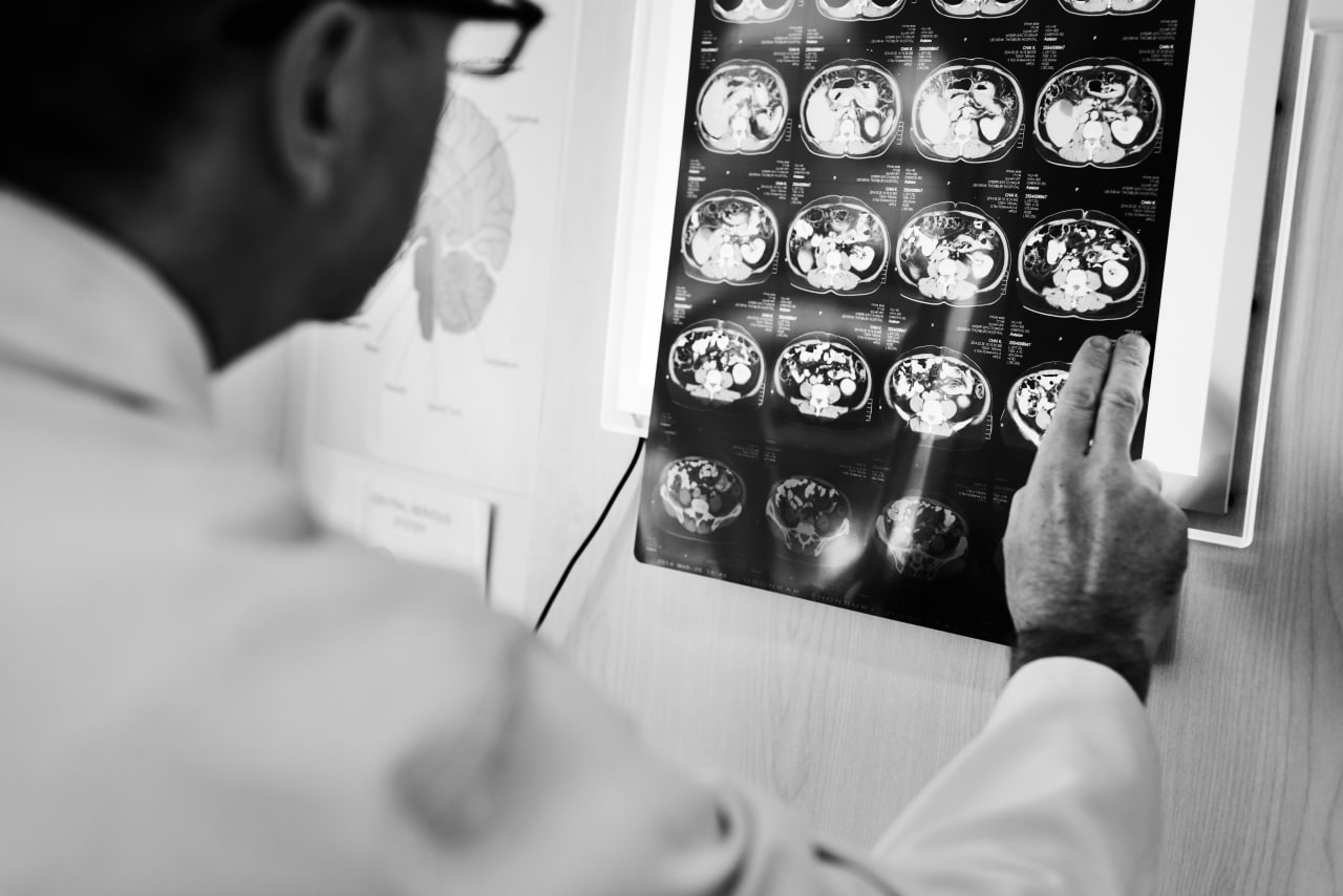
Image-guided surgery may be used for some brain tumours. Images are repeatedly taken with an MRI or a CT scan during the operation to show the location of the tumour and the surgeon’s instruments.
Once the surgeon has removed as much of the tumour as possible, the dura mater is tightly stitched together, the piece of skull is replaced with small screws and plates and the scalp is closed with stitches or staples. If the brain is very swollen after surgery, the piece of skull may be replaced later when the swelling has gone down. Healing usually takes several weeks.
- Brain Mapping
-
Brain mapping is done during a craniotomy when a tumour is near areas of the brain that control speech or motor function. Mapping is done with a technique called intraoperative cortical stimulation. It involves stimulating the surface of the brain with a mild electrical current to determine the function of a particular part of the brain. The procedure is painless. It produces temporary speech disturbances or twitching in the part of the body that is controlled by the area of the brain being stimulated. This information is then mapped so that the surgeon can avoid these areas when removing the tumour.
Speech mapping tracks the areas around the tumour that are responsible for speech and understanding speech. After a general anesthetic is given, the surgeon opens the skull and dura mater to expose the brain. You are then woken up so you can talk to the surgeon and follow instructions (such as counting or reading) during the mapping. When the mapping is finished, you are given a general anesthetic again and the surgeon continues the operation to remove the tumour.
Motor mapping tracks the areas around the tumour that are responsible for movement and reflexes. You may remain under general anesthetic. The surgeon stimulates the areas around the tumour with an electrical current and watches for any movement of the body. As with speech mapping, the surgeon uses the mapped areas as a guide when removing the brain tumour.